Anatomy Of The Upper Chest Area - Rahul's Medical Images: Medical Images » Chest Wall ... : Лучшие отзывы о курсе anatomy of the chest, abdomen, and pelvis.
Anatomy Of The Upper Chest Area - Rahul's Medical Images: Medical Images » Chest Wall ... : Лучшие отзывы о курсе anatomy of the chest, abdomen, and pelvis.. Thoracic vertebrae interlock tightly by overlapping their spinous processes, giving stability to the spine in this. The chest is part of a larger group of pushing muscles found in the upper body. Anatomy of the chest wall and breast. The anterior muscles of the trunk (torso) are associated with the front of the body, include chest and attachments: The trapezius originates from the skull and spine of the upper back and neck.
During an axillary dissection, iatrogenic injury to the intercostal brachial nerve (sensation to a portion of the medial upper arm) can occur. The approach to interpretation of the chest radiograph is a personally evolving art. The muscle consists of three parts which fan out during. Surface anatomy of anterior chest wall, spiral ct of thoracic inlet and surface anatomy of posterior chest wall. Read here everything about its anatomy.
Armor Plated Pecs! from www.bodybuilding.com It is a rare but serious condition, with the potential to cause vascular compromise of the upper limb. A diagram of the heart's valves. The forehead is referred to as the frontal region. Atlas of anatomy of the human body: It is a muscular organ around the size of a closed fist, and it sits in the chest, slightly to the left of share on pinterest. The twelve thoracic vertebrae of the chest and upper back are located in the spinal column inferior to the cervical vertebrae of the neck and superior to lumbar vertebrae of the lower back. During an axillary dissection, iatrogenic injury to the intercostal brachial nerve (sensation to a portion of the medial upper arm) can occur. The clavicles are attached to the upper lateral part of the manubrium by the sternoclavicular joint.
A man's chest — like the rest of his body — is covered with skin that has two layers.
The anterior muscles of the trunk (torso) are associated with the front of the body, include chest and attachments: Anatomy is to physiology as geography is to history: See more ideas about anatomy, upper limb anatomy, anatomy and physiology. Iv contrast may be injected into a vein in the patient's arm or hand. Anatomy of the physical exam6мин. Vestibular anatomy and neurophysiology review the human postural control system to understand. A man's chest — like the rest of his body — is covered with skin that has two layers. Read here everything about its anatomy. Related online courses on physioplus. The forehead is referred to as the frontal region. I will therefore split the chest up into three parts: The chest is part of a larger group of pushing muscles found in the upper body. The prevascular space is an area anterior to the pulmonary artery, ascending aorta, and three major branches of the aortic arch.
Read here everything about its anatomy. Arteries of the left foot. It is a muscular organ around the size of a closed fist, and it sits in the chest, slightly to the left of share on pinterest. Muscles forming the chest wall, which aid in respiration. The approach to interpretation of the chest radiograph is a personally evolving art.
Top 10 Strongest Muscles in The Body | Pouted.com from www.pouted.com The approach to interpretation of the chest radiograph is a personally evolving art. The anatomy of the sternum. Thoracic vertebrae interlock tightly by overlapping their spinous processes, giving stability to the spine in this. The forehead is referred to as the frontal region. Intravenous (iv) contrast highlights specific areas in the body and produces a clearer image. Arteries of the left foot. Vestibular anatomy and neurophysiology online course: The muscle consists of three parts which fan out during.
Read here everything about its anatomy.
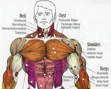
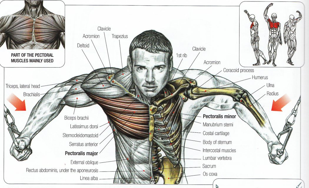
The chest anatomy includes the pectoralis major, pectoralis minor and the serratus anterior. Anatomy of the chest wall and breast. Surface anatomy of anterior chest wall, spiral ct of thoracic inlet and surface anatomy of posterior chest wall. Anatomy of the physical exam6мин. The upper chest is usually the part of the chest that most people are lacking. It forms the bulk of the chest area and can be easily seen on the surface in some people, for example weightlifters. Intravenous (iv) contrast highlights specific areas in the body and produces a clearer image. Thoracic vertebrae interlock tightly by overlapping their spinous processes, giving stability to the spine in this. Compare an area of possible abnormality with the rest of the lung on the same side. See more ideas about anatomy, upper limb anatomy, anatomy and physiology. Upper back pain and chest pain can occur together. Instant anatomy is a specialised web site for you to learn all about human anatomy of the body with diagrams, podcasts and revision questions. The cranial region encompasses the upper part of the head while the.
This illustration labeled regions of the human body show an anterior and posterior view of the body. Anatomy of the chest wall and breast. This area of the chest has attachments at the clavicle and the humerus or upper arm bone. A diagram of the heart's valves. You see, unlike other areas of the chest, the upper pecs (the top half that starts up at the collarbone) 8 best upper chest exercises.
Upper Chest Muscle Group - 6 Pack Abs Are Still Possible ... from i.pinimg.com Find out more about the individual muscles within the chest the chest is part of a larger group of pushing muscles found in the upper body. Iv contrast may be injected into a vein in the patient's arm or hand. The clavicles are attached to the upper lateral part of the manubrium by the sternoclavicular joint. Related online courses on physioplus. It forms the bulk of the chest area and can be easily seen on the surface in some people, for example weightlifters. The lungs are assessed and described by dividing them into upper, middle and lower zones. Find subtle abnormalities by using the sihouette sign. This illustration labeled regions of the human body show an anterior and posterior view of the body.
Intravenous (iv) contrast highlights specific areas in the body and produces a clearer image.
It describes the theatre of events. Iv contrast may be injected into a vein in the patient's arm or hand. I will therefore split the chest up into three parts: The cranial region encompasses the upper part of the head while the. Related online courses on physioplus. Read here everything about its anatomy. Arteries of the left foot. This illustration labeled regions of the human body show an anterior and posterior view of the body. Now that we've covered the anatomy and direction of the fibers. It is a muscular organ around the size of a closed fist, and it sits in the chest, slightly to the left of share on pinterest. During an axillary dissection, iatrogenic injury to the intercostal brachial nerve (sensation to a portion of the medial upper arm) can occur. The chest is part of a larger group of pushing muscles found in the upper body. Vestibular anatomy and neurophysiology online course: